Beta-thalassemia, a genetic blood disorder characterized by reduced or absent production of beta-globin chains, presents a fascinating array of skeletal manifestations that dramatically alter the skull’s structure and appearance. Beyond its well-known impact on blood cells, the disorder creates a distinctive set of radiological changes that tell a compelling story of the body’s adaptive response to chronic hemolytic anemia.
Beta-thalassemia is an inherited blood disorder that affects hemoglobin production. Patients with this condition experience chronic anemia due to the body’s inability to produce sufficient functional hemoglobin. To compensate for the reduced oxygen-carrying capacity of their blood, the body initiates a series of complex compensatory mechanisms that profoundly impact bone development, particularly in the skull.
The skull in beta-thalassemia patients undergoes several characteristic changes that are readily identifiable through medical imaging:
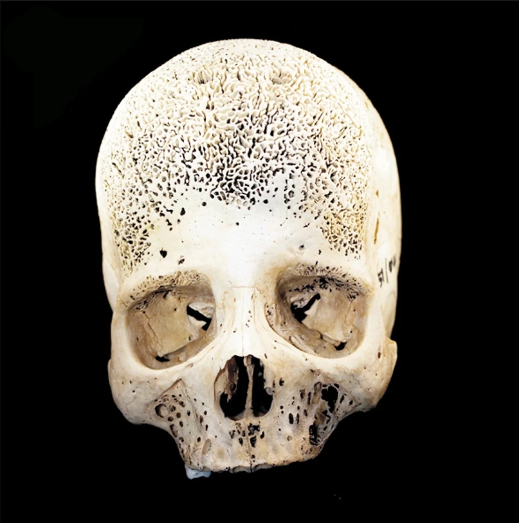
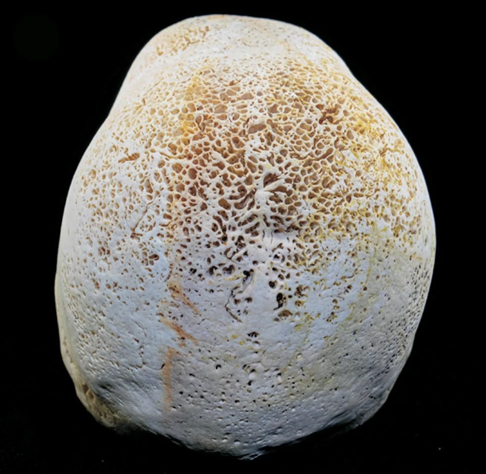
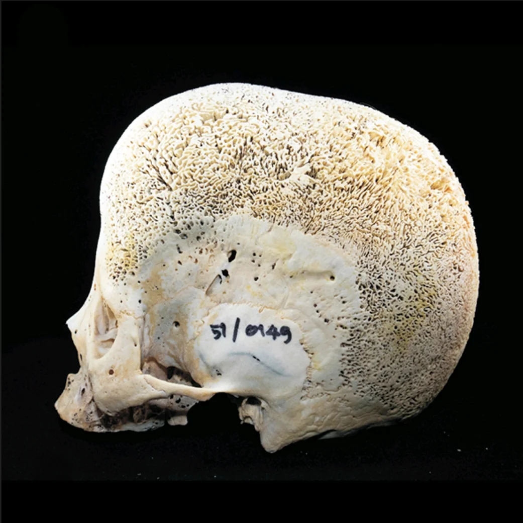
- Bone Marrow Hyperplasia The most significant transformation occurs in the bone marrow spaces. As the body attempts to increase red blood cell production, the bone marrow expands dramatically, causing distinctive radiological changes in the skull’s internal structure. This hyperplasia leads to notable alterations in the skull’s trabecular pattern and overall bone density.
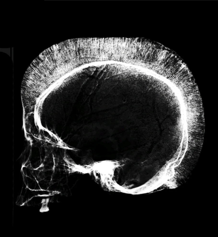
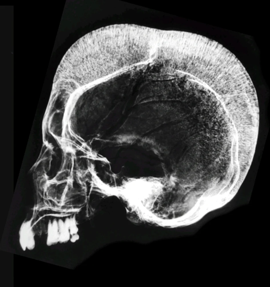
- Distinctive Skull Morphology Patients typically develop a distinctive “hair-on-end” appearance on radiographic images. This unique pattern resembles parallel hair-like striations running vertically across the skull, resulting from the compensatory bone marrow expansion and increased bone remodeling.
- Frontal and Parietal Bone Changes The frontal and parietal bones are particularly affected. They become markedly thicker and may show a characteristic “box-like” appearance. This dramatic remodeling is the body’s attempt to create additional space for expanded bone marrow and to support increased red blood cell production.
Children with beta-thalassemia often experience the most pronounced skull transformations during early development. The skull’s growth patterns become significantly altered, with:
- Increased skull diameter
- Prominence of the frontal and parietal regions
- Potential asymmetrical bone development
- Expanded diplöic spaces (the layer between the inner and outer skull tables)
These skull changes are more than just radiological curiosities. They serve as critical diagnostic indicators and provide insights into the disease’s progression and severity. Medical professionals use these characteristic changes to:
- Assess disease progression
- Monitor treatment effectiveness
- Predict potential complications
- Understand the patient’s overall physiological response
The skull transformations result from a complex interplay of physiological processes:
- Chronic hemolysis stimulates increased erythropoiesis (red blood cell production)
- Bone marrow expands to compensate for reduced hemoglobin
- Increased metabolic demands trigger extensive bone remodeling
- Compensatory mechanisms lead to characteristic skeletal changes
For patients, these skull changes can have significant psychological and social implications. The altered skull morphology may impact facial appearance, potentially affecting self-image and social interactions. Medical teams increasingly recognize the importance of comprehensive care that addresses both physical and emotional aspects of the condition.
Modern medical imaging has revolutionized our understanding of beta-thalassemia’s skeletal manifestations. Techniques such as:
- High-resolution CT scans
- 3D reconstruction imaging
- Advanced MRI protocols
Now provide unprecedented detailed views of the skull’s transformation, allowing for more precise monitoring and intervention.
Ongoing research continues to explore the intricate relationship between beta-thalassemia and skeletal development. Emerging studies focus on:
- Genetic mechanisms behind bone remodeling
- Potential targeted interventions
- Long-term skeletal health management strategies
The skull changes in beta-thalassemia represent a remarkable example of human physiological adaptation. They demonstrate the body’s extraordinary capacity to respond to chronic medical conditions, transforming bone structure in an attempt to maintain critical biological functions.
These radiological changes are more than medical anomalies—they are a testament to the complex, adaptive nature of human biology. Each altered skull tells a story of the body’s resilience, its constant struggle to maintain homeostasis in the face of genetic challenges.